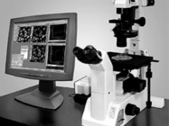
Scientific image analysis used to be performed by the human eye alone. Determining which spot was darker on a blot or what was going on in a cell culture dish has traditionally been as much art as science. The introduction of software for automated image analysis raised the bar by enabling much faster and more accurate analysis of laboratory images. When people hear the term 'image-analysis software,' they immediately think of images collected under a microscope,but the software can be used for just about any type of graphical data readout, including gels, fluorescence assays and even scans of organs orwhole animals. This article will address technology for cellular image-analysis software. There are a great many software packages to choose from for cellular image analysis. The specific intended application must be taken into account when considering products and features.
Open-source or commercial?
There are two major open-source products available for laboratory use—CellProfiler, from the Broad Institute of Massachusetts Institute of Technology and Harvard, and ImageJ,from the National Institutes of Health (NIH). They have gained a large following among users and offer an impressive menu of features and application-specific modules.
CellProfiler is designed to help biologists who lack technical computer-imaging skills obtain quantitative phenotypes from a large number of images. Its companion software, CellProfiler Analyst, is geared for the power user and permits interactive data analysis from high-throughput screens. CellProfiler’s machine-learning system can be trained to recognize specialized phenotypes and automatically score millions of images.
ImageJ was inspired by NIH Image for the Macintosh. It is a Java-based program that can be used online or downloaded and run on a computer. It reads a variety of image formats and supports a number of image-analysistasks, such as analyzing images in stacks, making measurements within images and manipulating images for better analysis. A recent improvementin CellProfiler has made it compatible with ImageJ.
“The full motivation for the [CellProfiler] project was to put sophisticated computational algorithms in the hands of biologists who may not have expertise to understand or write their own algorithm,” saysAnne Carpenter, Ph.D., a co-founder of the CellProfiler project at the Broad Institute.
Chris Kier, director of marketing for MetaMorph Software at Molecular Devicescalled CellProfiler “a nice package,” but adds, “the problem with all of the open-source packages is that today they don't seem to have the critical mass to keep up the support that's required by the end users.”
Martin Daffertshofer, global product manager, software, at PerkinElmer, says the open-source packages are “quite flexible” but adds that the user interface is “not optimal.”
For users who are self-sufficient and have facility with image analysis,open-source packages may be the only software they ever need. For thosewho want more support or don't have time to master the user interface offered by CellProfiler and ImageJ, a commercial product like MetaMorph or PerkinElmer's Volocity might be more accessible.
Matching solutions to applications
Different software packages have different features for specialized applications. For example, according to Carpenter, the CellProfiler teamis working to perfect algorithms that can identify different microscopic worms relative to each other and measure their properties. The worms stick to each other, sometimes overlapping, and have a more linear appearance than mammalian cell types. “Detangling of these worms—that's a bit of a challenge,” says Carpenter, “especially when they have a lot of small progeny.”
Another tangling problem, the tangling of neurites in neuronal cells, also confounds many software packages. Imaris, from Bitplane,excels at analyzing the types of tangled structures produced by neuronal cells, providing, for example, visualization of dendritic spines. Imaris “is the only software that can do this faithfully,” says Marcin Barszczewski, a product manager for Bitplane.
In addition to differing in performance within biological subspecialties, image-analysis software also varies according to speed of image acquisition and processing. Some software packages work very well for in-depth analysis of individual images, and others excel at high-throughput imaging.
CellProfiler and PerkinElmer's Columbus are examples of image-analysis software designed for high-throughput applications. According to PerkinElmer, the Columbus software uses a translocation script to analyze a 384-well plate, measured using an Opera QEHS with two fields per well and two channels per field, in less than 30 minutes.
Alternatively, some software is optimized for exhaustive analysis of single images. Imaris is powered to analyze image files of up to 50 gigabytes. “Some software packages I worked with in my research would [freeze] when images exceeded a few gigabytes,” says Barszczewski.
Innovative features
Today's image-analysis software includes some features never dreamed of in the early days. Media Cybernetics'new Live Tiling and Live EDF module for Image-Pro Plus 7.0 creates large, tiled images with no need for an automated stage. The module can use images collected manually by moving the stage around and stitching together the images collected into a single, larger image. It also can stitch together images collected through the Z-axis, creating a three-dimensional image that is fully in focus.
“No matter how deep the sample is, you can capture one in-focus image,” says Kathy Hrach, product manager for Media Cybernetics.
Barszczewski says Imaris also has an advanced new feature that enables it to segment and analyze cells' components, including vesicular objectsof various sizes that belong to functionally different populations. In addition, cell images now can be processed to visualize their membranes together with various pools of vesicles, nuclei and cytoplasm.
3D and 4D imaging
Imaging in three and four dimensions is a growing trend. Visualizing thecell in more dimensions is necessary to fully characterize some complexinteractions. Three dimensions plus time—basically a three-dimensional “movie”—provide the most comprehensive coverage. Such an analysis would once have been impossible for a personal computer, but newer technologies have made 3D and 4D analysis a reality.
PerkinElmer's Volocity is designed for high-speed acquisition of 3D images. Its 64-bit capability allows it to process very large files for asingle time point. After the images are acquired, Volocity Quantitationcan identify, measure and track objects in two, three or four dimensions; Volocity Restoration can de-convolve fluorescent images to improve quality and resolution, sharpening and removing haze.
“In a nutshell, it's one solution that brings everything you typically need when dealing with confocal images in one software package,” says Daffertshofer.
Compatibility
When purchasing image-analysis software, it is important to consider compatibility. Molecular Devices’ MetaMorph is one of the more “venerable” packages on the market, according to Kier, and hardware compatibility has been a special focus of its development. Instrument drivers can be tricky to develop for all the hardware customers might want to use the software with, but Molecular Devices has invested the resources to develop a very comprehensive library of drivers, making itssoftware compatible with all major brands of microscopes and CCD cameras. “Pretty much everything you might want to do with a microscope-imaging rig, you can do and control with MetaMorph Software,”says Kier. According to Molecular Devices, MetaMorph NX, the latest release, supports more drivers for components and devices than any of its competitors.
Media Cybernetics’ Image-Pro also supports a broad range of hardware, focusing mainly on light microscopes. Hrach says, however, that even if the software does not have a driver for a specific instrument, there's agood chance it can analyze the image output. “As long as we support that file format, we can open it in our product. Whether or not you can capture it in our software, we can open and analyze a very large number of file formats.”
Recently, scientists at the University of California, San Francisco, have developed an open-source alternative dubbed MicroManager,for controlling microscopes to acquire images. This software also supports a wide variety of hardware components, and it is compatible with ImageJ.
For many scientists, the choice between image-analysis software productsmay ultimately come down to budget. However, within each budget category it is worth considering how well each software package meets the needs of your specific application as well as its track record for the specific cells and phenotypes you are analyzing. After that, consider the features, hardware compatibility and user accessibility based on your own comfort level and experience. Many vendors are workingto make their interfaces as intuitive as possible and to reduce the number of mouse clicks per operation.

