使用三种成像照片对比可精确诊断早期卵巢癌(图)
通过将三种不相关的成像工具组成一台新的仪器,美国康涅狄格州和加利福尼亚州大学的科学家们开发了一种新的诊断技术,可以进行早期卵巢癌的诊断,并进行最大限度防止癌细胞扩散的手术,这将比目前标准的卵巢完全切除的手术要好得多。
由于卵巢癌临床早期无症状,鉴别其组织类型及良恶性相当困难,卵巢癌行剖腹探查术中发现肿瘤局限于卵巢的仅占30%,卵巢大多数已扩散到子宫双侧附件及盆腔各器官,所以卵巢癌在诊断上确是一大难题。卵巢癌是相对常见的病,大约有1.4%的女性会患上这种病。如果发现得早,90%的病人都能活下来;发现得迟,癌细胞扩散到卵巢,存活率就低于30%。
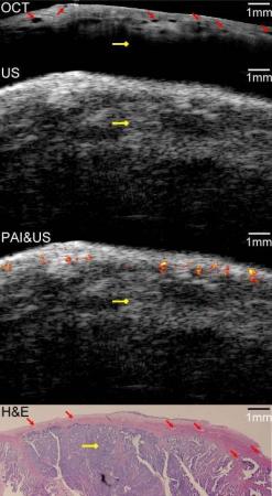
现在,使用这种全新的混合成像技术将能有效的进行卵巢癌的早期诊断。通过使用这台机器,科学家们能通过结合光声成像的对照照片、光学连续X线断层摄影术成像的高清表面照片、以及脉冲回波超声成像的组织深度成像照片来对卵巢癌进行诊断。他们在最新一期的《Biomedical Optics Express》杂志中详细描述了测试方法和结果。
科学家们使用了猪和人类卵巢组织进行了成像测试,发现能成功诊断出经后续组织染色和光镜检测才能确认的恶性肿瘤。虽然测试中使用的是手术移除的组织,但由于该设备的成像直径仅有5mm,因此在临床上完全可以通过切开一个小口并将其插入到组织中进行活检。 (生物探索 Jun)
生物探索推荐英文论文原文摘要:
Integrated optical coherence tomography, ultrasound and photoacoustic imaging for ovarian tissue characterization
Ovarian cancer has the lowest survival rate of the gynecologic cancers because it is predominantly diagnosed in Stages III or IV due to the lack of reliable symptoms, as well as the lack of efficacious screening techniques. Detection before the malignancy spreads or at the early stage would greatly improve the survival and benefit patient health. In this report, we present an integrated optical coherence tomography (OCT), ultrasound (US) and photoacoustic imaging (PAI) prototype endoscopy system for ovarian tissue characterization. The overall diameter of the prototype endoscope is 5 mm which is suitable for insertion through a standard 5-12.5mm endoscopic laparoscopic port during minimally invasive surgery. It consists of a ball-lensed OCT sample arm probe, a multimode fiber having the output end polished at 45 degree angle so as to deliver the light perpendicularly for PAI, and a high frequency ultrasound transducer with 35MHz center frequency. System characterizations of OCT, US and PAI are presented. In addition, results obtained from ex vivo porcine and human ovarian tissues are presented. The optical absorption contrast provided by PAI, the high resolution subsurface morphology provided by OCT, and the deeper tissue structure imaged by US demonstrate the synergy of the combined endoscopy and the superior performance of this hybrid device over each modality alone in ovarian tissue characterization.


