摘要:近日来自浙江大学生命研究院的管坤良教授、赵斌教授及加州大学圣地亚哥分校的Karen Tumaneng教授联合在国际著名期刊《自然—细胞生物学》(Nature cell biology)上发表了题为“The Hippo pathway in organ size control, tissue regeneration and stem cell self-renewal”的综述性文章。
这篇文章的通讯作者是浙江大学生命研究院管坤良教授,其早年毕业于杭州大学,现任职为美国加州大学圣地亚哥分校(UCSD)药学系教授,浙江大学生命科学研究院兼职教授、共同院长,复旦大学生物医学研究院基因组研究所和分子细胞生物学实验室兼职杰出PI。管坤良教授长期以来主要从事细胞生长调控、肿瘤生物学以及神经生物学的信号转导途径等方面的研究。以其突出的科研成果曾荣获美国“麦克阿瑟天才奖”、MacArthur基金颁发的MacArthur大奖、美国生物化学和分子生物学学会“青年科学家奖”和北美华人生物学会“青年科学奖”等,多次在 Cell, Nature, Science, Genes & Development, Nature Cell Biology 等国际顶级学术期刊发表论文。
生物体中组织器官大小和体积的调控一直是生物学研究的最基本的问题之一。Hippo信号通路是一条细胞抑制生长性信号通路,在进化过程中非常保守,多细胞动物果蝇、小鼠、哺乳动物中都存在Hippo信号通路。最早研究人员在果蝇中发现Hippo信号传导路径是调节细胞大小和器官体积的主要信号通路。之后研究人员证实Hippo信号通路具有高度保守型,果蝇中Hippo信号通路的成员都能在高等生物中找到对应的同源物。在哺乳动物中,Hippo信号通路上游的膜蛋白受体感受到胞外的生长抑制信号后,经过一系列激酶复合物的磷酸化级联反应,最终将磷酸化下游的效应因子YAP。磷酸化的YAP与细胞骨架蛋白相互作用,被滞留在胞质内,不能进入细胞核行使其转录激活功能,从而实现对器官大小和体积的调控。此外,近年来的研究证实Hippo信号传导通路还在癌症发生、组织再生以及干细胞的功能调控上发挥重要的作用。
在这篇综述性文章中,管坤良教授等基于其课题组的相关系列成果,结合近年来在相关领域的研究成果和最新进展,系统综述了Hippo信号传导通路组成及调控机制,并重点对器官大小和组织再生调控过程中细胞极性蛋白和细胞粘附蛋白对Hippo信号传导通路的影响及机制进行了综述和探讨。
近年来关于Hippo信号传导通路的研究十分活跃,处于国际科技发展的前沿。浙江大学生命科学研究院的多个课题组对于调控Hippo信号传导通路同时在展开深入性的研究。不久前浙江大学生命科学研究院的黄俊教授在对hippo信号通路关键蛋白:YAP1的研究中取得突破性进展。研究人员克隆出了两个YAP1结合蛋白AMOTL1和AMOTL2,并发现它们通过影响YAP1的核质穿梭副调控YAP的功能。进一步的研究发现,在MCF10A细胞中降低AMOTL2的表达能够促进细胞发生上皮细胞向间充质细胞的转化(EMT),而这一表型恰好与YAP1在该细胞中过量表达的表型一致。这一研究成果发表在美国生化与分子生物学学会主办的JBC杂志上。
生物探索推荐英文论文摘要:
The Hippo pathway in organ size control, tissue regeneration and stem cell self-renewal
Abstract:
Precise control of organ size is crucial during animal development and regeneration. In Drosophila and mammals, studies over the past decade have uncovered a critical role for the Hippo tumour-suppressor pathway in the regulation of organ size. Dysregulation of this pathway leads to massive overgrowth of tissue. The Hippo signalling pathway is highly conserved and limits organ size by phosphorylating and inhibiting the transcription co-activators YAP and TAZ in mammals and Yki in Drosophila, key regulators of proliferation and apoptosis. The Hippo pathway also has a critical role in the self-renewal and expansion of stem cells and tissue-specific progenitor cells, and has important functions in tissue regeneration. Emerging evidence shows that the Hippo pathway is regulated by cell polarity, cell adhesion and cell junction proteins. In this review we summarize current understanding of the composition and regulation of the Hippo pathway, and discuss how cell polarity and cell adhesion proteins inform the role of this pathway in organ size control and regeneration.
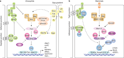
Figure 1: The Hippo pathway in Drosophila and mammals.
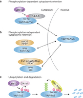
Figure 2: Mechanisms of YAP/TAZ/Yki inhibition by the Hippo pathway.
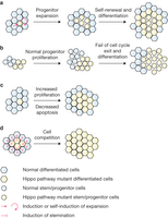
Figure 3: Mechanisms of the Hippo pathway in regulation of organ size and regeneration.


