摘要:据7月20日刊《美国医学会杂志》上的一则研究显示,相比过去的研究报告,应激性心肌病(这是一种短暂形式的心力衰竭,由应激事件所触发)或许有更广泛的临床特征,其中包括它会累及更年轻的患者、男性以及没有可被识别的应激触发因子等。
据文章的背景资料披露,应激性心肌病(SC)主要影响的是绝经后妇女,其特征为急性、深度,但可逆转的左心室(LV)功能障碍,而患者没有明显的冠心病。“该病临床特点的不同方面曾经在小型的单一中心的人群中被描述过,但是较大型、多中心的成组数据迄今为止则一直缺乏。此外,患者在入院时,应激性心肌病(SC)被快速诊断出来仍然很困难。”
德国莱比锡大学的Ingo Eitel, M.D.及其同事开展了一项研究,旨在全面定义SC的临床谱,并对疑似SC的诊断决定可能有帮助的一组心血管磁共振(CMR)标准的可用性进行了检验。这项研究是在欧洲和北美的7家三级医疗中心进行的,时间是2005年1月至2010年10月间。研究人员对256名SC患者在各中心期间以及在急性发病之后1个月至6个月时的情况进行了评估。
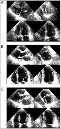
患有SC的病人的平均年龄为69岁;89%(n = 227)的病人为女性。81%的病人(n = 207)为绝经后妇女;有20名妇女(占8%)的年龄为50岁或以下;男性占了11%的病例。71%的病人(n = 182)在发病前不到48小时中存在有一个明显的应激事件;触发条件为情绪压力(30%)及生理压力(41%)。当患者来到医疗中心的时候,87%的患者在心电图(ECGs)上显示有异常。冠状动脉造影显示,有193名患者(占75%)有着健康的冠状动脉。CMR成像发现有气球样模式(这是心肌的一种形貌)及在所有病人中都有中度至重度的LV功能减损及4种独特的区域性心室气球样模式。
文章的作者写道:“人们可用特定的标准由CMR精确地识别应激性心肌病:一种典型的LV功能障碍模式、心肌水肿、没有明显的坏死(细胞或组织的死亡)/纤维化(过多纤维性结缔组织的形成)以及心肌炎症的标记。追踪性CMR成像显示,所有患者在没有明显纤维化的情况下,LV射血分数(这是对心脏每次收缩时左心室泵血功能的一种测定方法)和炎症标记完全恢复正常。”
研究人员指出,他们的数据表明,只有三分之二的病人有着清楚可辨的前发性应激因子,然而在以往的报告中,具有前发性情绪或生理触发因子的百分比高达89%。 “因此,我们的大型多中心的群组证明,没有可辨识的应激性事件并不能排除这一诊断;所以该病的加剧机制可能更为复杂,例如它可能会牵涉到血管、内分泌及中枢神经系统。这一临床异质性可能与人们在认识SC上的不确定性有关,并因此可能会影响到该疾病的处理策略。因此,提高SC广泛临床特征的意识和认识,对疑似SC的患者中作出正确的诊断和治疗将是必须的。”(生物探索编辑)
应激性心肌病(Stress cardiomyopathy )
生物探索推荐英文论文摘要:
Clinical Characteristics and Cardiovascular Magnetic Resonance Findings in Stress (Takotsubo) Cardiomyopathy
ABSTRACT
Context Stress cardiomyopathy (SC) is a transient form of acute heart failure triggered by stressful events and associated with a distinctive left ventricular (LV) contraction pattern. Various aspects of its clinical profile have been described in small single-center populations, but larger, multicenter data sets have been lacking so far. Furthermore, it remains difficult to quickly establish diagnosis on admission.
Objectives To comprehensively define the clinical spectrum and evolution of SC in a large population, including tissue characterization data from cardiovascular magnetic resonance (CMR) imaging; and to establish a set of CMR criteria suitable for diagnostic decision making in patients acutely presenting with suspected SC.
Design, Setting, and Patients Prospective study conducted at 7 tertiary care centers in Europe and North America between January 2005 and October 2010 among 256 patients with SC assessed at the time of presentation as well as 1 to 6 months after the acute event.
Main Outcome Measures Complete recovery of LV dysfunction.
Results Eighty-one percent of patients (n = 207) were postmenopausal women, 8% (n = 20) were younger women (aged ≤50 years), and 11% (n = 29) were men. A stressful trigger could be identified in 182 patients (71%). Cardiovascular magnetic resonance imaging data (available for 239 patients [93%]) revealed 4 distinct patterns of regional ventricular ballooning: apical (n = 197 [82%]), biventricular (n = 81 [34%]), midventricular (n = 40 [17%]), and basal (n = 2 [1%]). Left ventricular ejection fraction was reduced (48% [SD, 11%]; 95% confidence interval [CI], 47%-50%) in all patients. Stress cardiomyopathy was accurately identified by CMR using specific criteria: a typical pattern of LV dysfunction, myocardial edema, absence of significant necrosis/fibrosis, and markers for myocardial inflammation. Follow-up CMR imaging showed complete normalization of LV ejection fraction (66% [SD, 7%]; 95% CI, 64%-68%) and inflammatory markers in the absence of significant fibrosis in all patients.
Conclusions The clinical profile of SC is considerably broader than reported previously. Cardiovascular magnetic resonance imaging at the time of initial clinical presentation may provide relevant functional and tissue information that might aid in the establishment of the diagnosis of SC.
KEYWORDS: coronary angiography, diagnosis, magnetic resonance imaging, stress, physiological, stress, psychological, takotsubo cardiomyopathy.


