英国科学家近日绘制出了首个全新的大脑3D成像技术
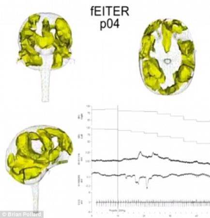
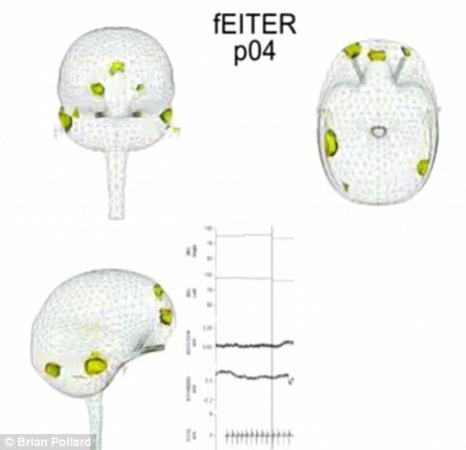
该3D成像技术是由一台机器来运行的,该机器为手提式的轻质量监视器,它甚至可以安置在一个小型手推车上,监视器上还有安置在病人脑部的32个电极(图)
据英国《每日邮报》报道,近几年,无论是游戏还是电影,只要加入3D技术,都颇受欢迎。医学界也不落后,英国科学家近日绘制出了首个全新的大脑3D成像技术,用3D视频清晰的记录了麻醉剂药物注射进大脑后,脑部从活跃到失去知觉的一系列变化,其中还可以观察到大脑深度脑电活动的变化过程。
英国一支研究小组使用了一个名为“能唤起回应的功能性电阻抗X线断层摄影术”的新3D成像方法来进行试验。该技术不仅能够高速成像,还能监视大脑中的深度脑电活跃度,“功能性电阻抗X线断层摄影术”给大脑带来的刺激,与麻醉剂等外界条件带来的刺激相同,从而实现科学家对大脑功能的准确测量。据介绍,该3D成像技术是由一台机器来运行的,该机器为手提式的轻质量监视器,它甚至可以安置在一个小型手推车上,监视器上还有安置在病人脑部的32个电极。当一股小且高频率的电流流过时,即使人们不能产生任何感觉,只要经过两个电极之间,就会测量到另外一组电极中产生的电压,整个过程用时间不到千分之一秒,随后,“电子扫描”就会开始运作。这台监视器在一秒钟内可以循环执行整个过程100次之多。通过测量大脑对电流的抵抗程度,大脑导电率变化的代表性图像就制作而成了。这个试验也会揭示出大脑不同部分的电波活跃程度。波勒德教授在本周的阿姆斯特丹欧洲麻醉学大会上表示,我们的研究发现说明,大脑无意识是由于被抑制的神经集合大规模遍布大脑皮层。
科学家在观看该大脑3D视频后发现,人体在被注入麻醉剂后,大脑中某组特定神经细胞的活跃性将会被改变,且大脑不同部分之间的联系也会被阻碍,这就导致了大脑最终失去知觉。
曼彻斯特大学的研究负责人布莱恩—波勒德(Brian Pollard)表示,这次的发现支持了英国牛津大学苏珊—格林菲尔德(Susan Greenfield)曾提出的关于意识本身性质的一种假设。格林菲尔德教授曾表示,大脑知觉是由不同组大脑细胞所组成的,这些细胞在一起高效工作,或者依赖于有效的感觉刺激而起作用,因此意识并不是处于一个全有或全无的状态,而更像一个调光器开关,根据人们的成长,情绪,或者药物来相应的做出变化。也就是说,一旦人们被麻醉后,大脑中的小型神经集合彼此就不会很好的工作,或者会与其他的神经集合断开联系。波勒德教授表示,虽然我们现在仍然不能完全了解当大脑处于无意识状态时,大脑内部究竟发生了什么。但这个发现却可以让我们对大脑及其功能的了解更加深入。
生物探索推荐英文原文报道:
Sit back and relax: Watch a 3-D film of the brain as it loses consciousness
For the first time researchers have watched the brain as it loses consciousness.
A new imaging method constructs a 3-D movie of the brain as it changes while an anaesthetic drug takes effect.
The real-time 3-D images show that slipping into unconsciousness involves a change in electrical activity deep within the brain, scientists said.
The shift is thought to alter the activity of certain groups of nerve cells and hinders communication between different parts of the brain.
Lead researcher Brian Pollard, of the University of Manchester, said the findings appear to support a hypothesis put forward by Professor Susan Greenfield, of Oxford University, about the nature of consciousness itself.
Professor Greenfield suggests consciousness is formed by different groups of brain cells, which work efficiently together, or not, depending on the available sensory stimulations, and that consciousness is not an all-or-none state but more like a dimmer switch, changing according to growth, mood or drugs.
When someone is anaesthetised it appears that small neural assemblies either work less well together, or inhibit communication with other neural assemblies.
Professor Pollard this week told the European Anaesthesiology Congress in Amsterdam: 'Our findings suggest that unconsciousness may be the increase of inhibitory assemblies across the brain's cortex.
'These findings lend support to Greenfield's hypothesis of neural assemblies forming consciousness.'
The team used a new imaging method called 'functional electrical impedance tomography by evoked response', or fEITER.
It enables high-speed imaging and monitoring of electrical activity deep within the brain and is designed to enable researchers to measure brain function.
The machine itself is a portable, light-weight monitor, which can fit on a small trolley. It has 32 electrodes that are fitted around the patient's head.
A small, high-frequency electric current - too small to be felt or have any effect - is passed between two of the electrodes, and the voltages between other pairs of electrodes are measured in a process that takes less than one thousandth of a second.
An 'electronic scan' is thus carried out and the machine does this whole procedure 100 times a second.
By measuring the resistance to current flow, a cross-sectional image of the changing electrical conductivity within the brain is constructed. This is thought to reflect the amount of electrical activity in different parts of the brain.
The speed of the response of fEITER is such that the evoked response of the brain to external stimuli, such as an anaesthetic drug, can be captured in rapid succession as different parts of the brain respond, thus tracking the brain's processing activity.
Professor Pollard said: 'We have looked at 20 healthy volunteers and are now looking at 20 anaesthetised patients scheduled for surgery.
'We are able to see 3-D images of the brain's conductivity change, and those where the patient is becoming anaesthetised are most interesting.
'We have been able to see a real-time loss of consciousness in anatomically distinct regions of the brain for the first time. We are currently working on trying to interpret the changes that we have observed.
'We still do not know exactly what happens within the brain as unconsciousness occurs, but this is another step in the direction of understanding the brain and its functions.'
Professor Pollard said that a huge amount of research still needed to be done to fully understand the role EIT could play in medicine.
He added: 'If its power can be harnessed, then it has the potential to make a huge impact on many areas of imaging in medicine.
'It should help us to better understand anaesthesia, sedation and unconsciousness, although its place in medicine is more likely to be in diagnosing changes to the brain that occur as a result of, for example, head injury, stroke and dementia.
'The biggest hurdle is working out what we are seeing and exactly what it means, and this will be an ongoing challenge.'


