摘要:为了增加临床实践的安全性和减少手术中可能出现的风险和损失,计算机和数字医疗影像是至关重要的。脑部手术前,神经外科医生可以评估患者手术的风险,从而提高安全性,避免一些不必要的风险。
This is the Fraunhofer MEVIS neuro exhibit.
Brain interventions must be planned so that the neurosurgeon can access and remove the tumor without causing unnecessary damage. Before the brain tumor can be removed, crucial questions must be answered. Where do the functional areas of the cortex (gray matter) of the patient lie? What are the paths of the nerve fiber tracts that connect them? Answering these questions is important because the functional areas of the brain are interconnected via nerve pathways, also known as nerve fiber tracts. These nerve tracts must be protected as much as possible; otherwise, permanent dysfunction could occur. Furthermore, nerve tracts can be pushed or infiltrated by the brain tumor itself. If nerve tracts become damaged during an operation, there is a risk that distant functional areas connected to the tumor-afflicted part of the brain could be affected and induce lasting sensory, motor, and cognitive impairment. Therefore, neurosurgeons attempt to answer these questions for each patient during the planning stage of the brain operation to minimize the risks present in the intervention. To do so, surgeons require medical imagery of each patient's brain anatomy and function that is as realistic and precise as possible. However, medical images contain inaccuracies that arise from the processing, modeling, and reconstruction of patient data.
Solving these problems requires more than merely improving existing imaging methods. Mathematical analysis and models must be integrated to produce information about the location of the tumor, functional areas, and nerve fiber tracts, to increase the accuracy of patient-specific data, and to give the surgeon dependable knowledge.
The Fraunhofer MEVIS Institute for Medical Image Computing in Bremen, Germany has pioneered a procedure that analyzes uncertainty in patient-specific images, modeling, and reconstruction and incorporates this information into reconstructions of patient data. This procedure allows safety margins around nerve tracts in the brain to be more accurately determined. In addition, the reliability of the reconstructed data is calculated to supply the surgeon with accurate information concerning nerve tract locations, paths, and intersections and to construct safety margins around the nerve fiber tracts. By integrating errors in measurement, reconstruction, and modeling, the exact locations of tracts in a space-occupying tumor are calculated. This gives the neurosurgeon a reliable prognosis concerning where the incision in the brain should be made and which safety margins should be chosen to avoid harming nerve tracts and irreversibly damaging important functional areas. Before an intervention, the surgeon can evaluate patient-specific risks. These software assistants will be refined and implemented for neuronavigation in future operations, providing the surgeon with updated information during surgery that can be compared to planning data.
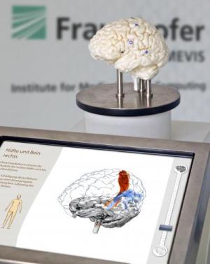
The paths of nerve tracts in the brain and the functional areas that they connect can now be explored by visitors of the "New Paths in Medicine" exhibit on the MS Wissenschaft exhibition ship. The converted inland vessel is underway until September 29, 2011 and docks in 35 different cities. During the "Year of Health Research," visitors can familiarize themselves with the field's newest trends, developments, and research findings. The exhibit showcases a physical three-dimensional model of the brain produced through an innovative printing process based on the medical image data of a real person. This brain model can be touched and viewed from different angles thanks to its rotating base. Nerve tracts can be activated by touching sensors on the physical model that correspond to functional areas of the brain. The brain is displayed on a screen along with the activated nerve tracts that are responsible, for instance, for sight, speech, feeling, and motion. This new form of interactive exhibit was developed by Fraunhofer MEVIS in Bremen together with the Universum® Science Center in Bremen to demonstrate how modern image processing combined with mathematics and intelligent software can help make neurosurgical operations more predictable and safe. The three-dimensional print of the brain was produced by the Fraunhofer-Institut ITWM in Kaiserslautern.


