生物谷Bioon.com 讯 8月6日,重庆大学生物工程学院邓林红教授实验室与美国哈佛大学合作实验室在PLoS ONE上发表论文,揭示机械拉伸引发细胞流态化响应的内在机制。
该合作研究是邓林红教授与哈佛大学合作者继2007年在Nature上发表有关机械拉伸引起细胞流态化响应论文后的进一步深化。(Xavier Trepat,Linhong Deng et al.Universal physical responses to stretch in the living cell.Nature. doi:10.1038/nature05824)。2007年发表在Nature上的论文首次提出了活细胞在收到短暂机械牵张之后会迅速软化并向流体状态转化,随后又逐渐恢复到拉伸前的状态。这一现象在各种细胞和生化环境下具有普遍性,因此对于理解细胞的物理行为和相关生理病理现象具有十分重要的意义。但该现象的内在机制仍然没有得到完全的揭示。
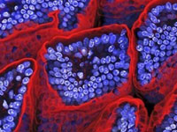
该文章第一作者陈诚等利用细胞牵张力显微测量技术,磁微粒扭转细胞流变测量技术等细胞力学领域先进技术动态观测了人体膀胱平滑肌细胞在受到短暂的机械牵张后,其细胞硬度、细胞牵张力、以及细胞骨架形态的变化。实验结果表明,膀胱平滑肌细胞在受到短暂牵张之后迅速"液化",然后缓慢的"再固化",逐渐恢复到拉伸前的状态。更为重要的是,由于短暂拉伸持续的时间非常短,细胞发生的所有物理相应过程并不是通过细胞内部的生物化学信号通路调控的,而是通过细胞骨架的纤维型肌动蛋白(F-actin)的迅速分解和缓慢再重组来实现和调控的。
论文不仅采用人体内另一种同样收到机械牵张力的影响的膀胱平滑肌细胞进一步证实了细胞在受到短暂拉伸后迅速发生流态化与缓慢恢复的普遍性,并且找出首次观测到了F-actin在其中扮演的关键作用,部分揭示了此现象的内在物理调控机制。
随着现代细胞生物学和细胞动力学的共同发展,机械力和物理环境如何对细胞结构和功能发生影响并决定许多重大生命活动过程的谜团正在被逐步打开。因此,在对细胞进行传统的生物分析和化学分析的同时,系统地研究细胞的物理环境和受力情况以及相应的细胞行为规律,将对更准确、更深入地理解细胞的运作机制、为细胞生理学和病理学的研究提供新的启发和思路。
生物谷推荐英文摘要:
PLoS ONE doi:10.1371/journal.pone.0012035
Fluidization and Resolidification of the Human Bladder Smooth Muscle Cell in Response to Transient Stretch
Cheng Chen1,2#, Ramaswamy Krishnan2#, Enhua Zhou2, Aruna Ramachandran3, Dhananjay Tambe2, Kavitha Rajendran2, Rosalyn M. Adam3, Linhong Deng1,2*, Jeffrey J. Fredberg2
1 Key Laboratory of Biorheological Science and Technology, Ministry of Education, Bioengineering College, Chongqing University, Chongqing, China, 2 Program in Molecular and Integrative Physiological Sciences, Department of Environmental Health, Harvard School of Public Health, Boston, Massachusetts, United States of America, 3 Urological Diseases Research Center, Department of Urology, Children's Hospital Boston and Department of Surgery, Harvard Medical School, Boston, Massachusetts, United States of America
Abstract
Background:Cells resident in certain hollow organs are subjected routinely to large transient stretches, including every adherent cell resident in lungs, heart, great vessels, gut, and bladder. We have shown recently that in response to a transient stretch the adherent eukaryotic cell promptly fluidizes and then gradually resolidifies, but mechanism is not yet understood.
Principal Findings:In the isolated human bladder smooth muscle cell, here we applied a 10% transient stretch while measuring cell traction forces, elastic modulus, F-actin imaging and the F-actin/G-actin ratio. Immediately after a transient stretch, F-actin levels and cell stiffness were lower by about 50%, and traction forces were lower by about 70%, both indicative of prompt fluidization. Within 5min, F-actin levels recovered completely, cell stiffness recovered by about 90%, and traction forces recovered by about 60%, all indicative of resolidification. The extent of the fluidization response was uninfluenced by a variety of signaling inhibitors, and, surprisingly, was localized to the unstretch phase of the stretch-unstretch maneuver in a manner suggestive of cytoskeletal catch bonds. When we applied an "unstretch-restretch" (transient compression), rather than a "stretch-unstretch" (transient stretch), the cell did not fluidize and the actin network did not depolymerize.
Conclusions:Taken together, these results implicate extremely rapid actin disassembly in the fluidization response, and slow actin reassembly in the resolidification response. In the bladder smooth muscle cell, the fluidization response to transient stretch occurs not through signaling pathways, but rather through release of increased tensile forces that drive acute disassociation of actin.


