导读:卡尔斯鲁厄理工学院和海德堡大学的研究人员使用了一种新颖的高分辨率成像方法,首次成功地实时观察活体中细胞膜的修复过程。研究结果有助于人类肌肉疾病治疗的发展以及为生物技术开发出更多发展潜力。

每个细胞都由薄的脂质双层所包围,从而隔离开细胞独特的内环境与细胞外环境。破坏脂质双层也就是细胞膜会破坏细胞功能,甚至可能导致细胞的死亡。举例来说,走下坡路会使我们腿部肌肉细胞的细胞膜出现许多小孔。为了避免不可修复的伤害,肌肉细胞有高效的系统可以再次密封这些小孔。
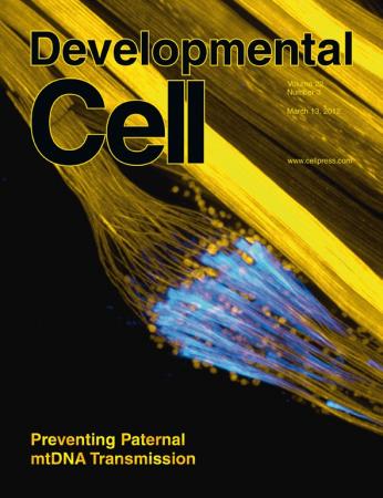
Uwe Strähle 和Urmas Roostalu博士使用了一种新颖的高分辨率成像方法,首次实时观察了活体动物的细胞膜修复过程。他们用荧光蛋白标记了斑马鱼幼体肌肉组织的修复蛋白。研究人员用激光照射肌肉细胞的细胞膜并烧出一些小孔,然后在显微镜下实时地观察这些小孔的修复过程。观察发现膜小泡和两种蛋白质Dysferlin和膜联蛋白A6快速地形成修复补丁。其他的膜联蛋白随后积聚在受伤的细胞膜处。卡尔斯鲁厄和海德尔堡的研究人员发现,细胞从内部积聚了多层修复补丁在细胞外坏境中密封细胞内部。此外,研究人员还发现存在一个特定的细胞膜区域,可以快速地为细胞膜提供修复膜上小孔的物质。
膜修复的动物模型会继续确定在密封过程新的蛋白质,并且帮助阐明这其中的机理。研究结果有助于人类肌肉疾病治疗的发展以及为生物技术开发出更多发展潜力。

In Vivo Imaging of Molecular Interactions at Damaged Sarcolemma
Urmas Roostalu, Uwe Strähle
Muscle cells have a remarkable capability to repair plasma membrane lesions. Mutations in dysferlin (dysf) are known to elicit a progressive myopathy in humans, probably due to impaired sarcolemmal repair. We show here that loss of Dysf and annexin A6 (Anxa6) function lead to myopathy in zebrafish. By use of high-resolution imaging of myofibers in intact animals, we reveal sequential phases in sarcolemmal repair. Initially, membrane vesicles enriched in Dysf together with cytoplasmic Anxa6 form a tight patch at the lesion independently of one another. In the subsequent steps, annexin A2a (Anxa2a) followed by annexin A1a (Anxa1a) accumulate at the patch; the recruitment of these annexins depends on Dysf and Anxa6. Thus, sarcolemmal repair relies on the ordered assembly of a protein-membrane scaffold. Moreover, we provide several lines of evidence that the membrane for sarcolemmal repair is derived from a specialized plasma membrane compartment.
文献链接:https://www.physorg.com/news/2012-03-muscle-cells-membranes.html

