导读:美国研究人员13日公布的一项研究报告显示,长时间太空飞行可能导致宇航员出现大脑和视觉受损,其症状与不明原因的颅内高压患者类似。

研究人员对27名在航天飞机和国际空间站内的微重力或零重力环境中平均度过108天的宇航员进行了核磁共振成像分析。他们发现,9名宇航员视神经周围的脑脊液空间有所扩大,6名宇航员眼球后部变得扁平,4人视神经肿胀,3人的脑垂体及其与大脑的连接发生变化。这些症状常见于一些不明原因的颅内高压患者当中,颅内压力升高可导致视神经和眼球的节点膨胀,损害视力。
发生变化的脑垂体也引起研究者关注。脑垂体是人体内最复杂、最重要的内分泌腺,能分泌多种对代谢、生长、发育和生殖至关重要的激素。
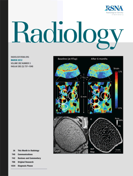
这项研究当天发表在美国期刊《放射学》网络版上。领导该研究的得克萨斯大学医学院教授拉里·克雷默表示,核磁共振成像揭示了宇航员暴露于微重力环境后出现的多种异常,这也有助于医学专家更好地理解颅内高压的形成机制。
此前有研究表明,长期太空生活可导致宇航员骨质疏松、肌肉和视力退化。例如国际空间站的宇航员在度过6个月失重生活并重返地面后,往往需要一年多时间才能恢复原有骨质。
美国航天局约翰逊航天中心官员威廉·塔弗13日在一份声明中表示,美航天局已经注意到部分宇航员的视力有所变化,其出现颅内高压的可能性虽然存在但仍有不确定性,美航天局将开始研究这些情况背后的机制并密切监控。

Orbital and Intracranial Effects of Microgravity: Findings at 3-T MR Imaging
Larry A. Kramer, MD, Ashot E. Sargsyan, MD, Khader M. Hasan, PhD, James D. Polk, DO and Douglas R. Hamilton, MD, PhD1
Purpose: To identify intraorbital and intracranial abnormalities in astronauts previously exposed to microgravity by using quantitative and qualitative magnetic resonance (MR) techniques.
Materials and Methods: The institutional review board approved this HIPAA-compliant, retrospective review and waived the requirement for informed consent. Twenty-seven astronauts (mean age ± standard deviation, 48 years ± 4.5) underwent 3-T MR imaging with use of thin-section, three-dimensional, axial T2-weighted orbital and conventional brain sequences. Eight astronauts underwent repeat imaging after an additional mission in space. Optic nerve sheath diameter (ONSD) and optic nerve diameter (OND) were quantified in the retrolaminar optic nerve. OND and central optic nerve T2 hyperintensity were quantified at mid orbit. Qualitative analysis of the optic nerve sheath, optic disc, posterior globe, and pituitary gland morphology was performed and correlated for association with intracranial evidence of hydrocephalus, vasogenic edema, central venous thrombosis, and/or mass lesion. Statistical analyses included the paired t test, Mann-Whitney nonparametric test for group comparisons, Cronbach α coefficient for reproducibility, and Pearson correlation coefficient.
Results: All astronauts had previous exposure to microgravity and, thus, control data were not available for comparison. The ONSD and OND ranged from 4.7 to 10.8 mm (mean, 6.2 mm ± 1.1) and from 2.4 to 4.5 mm (mean, 3.0 mm ± 0.5), respectively. Posterior globe flattening was seen in seven of the 27 astronauts (26%), optic nerve protrusion in four (15%), and moderate concavity of the pituitary dome with posterior stalk deviation in three (11%) without additional intracranial abnormalities. Retrolaminar OND increased linearly relative to ONSD (r = 0.797, Pearson correlation). A central area of T2 hyperintensity was identifiable in 26 of the 27 astronauts (96%) and increased in diameter in association with kinking of the optic nerve sheath.
Conclusion: Exposure to microgravity can result in a spectrum of intraorbital and intracranial findings similar to those in idiopathic intracranial hypertension.
文献链接:https://radiology.rsna.org/content/early/2012/03/07/radiol.12111986.abstract

