中科院西安光机所瞬态光学与光子技术国家重点实验室姚保利研究组,将基于数字微镜器件和LED照明的显微技术成功用于生物医学研究,从而为深层生物样品大面积快速三维成像提供了一种新的技术手段。相关成果日前发表在《自然》子刊《科学报告》杂志上。
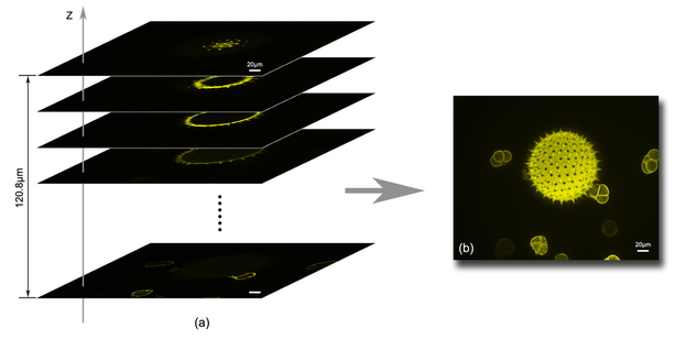
姚保利研究组利用新方法得到的植物花粉三维光切片成像
大到宇宙,小到分子,看得更远、更细、更清楚是人类不断追求的目标。为突破光的衍射极限,近年来出现了不少光学超分辨方法,如光激活定位法、随机光学重构法、受激发射损耗法等。但这几种超分辨成像技术速度较慢,而且需要一些特殊染料标记样品。另外一种方式是使用结构光照明的显微技术(SIM)。它使用特殊调制的光场照明样品,通过空间频谱处理的方式获得超分辨图像。目前,只有美、德、英、日等几个国家掌握该技术,我国在这方面相对滞后。
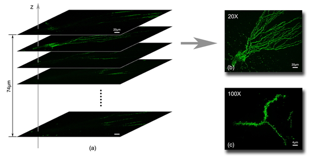
姚保利研究组利用新方法得到的小鼠脑神经元细胞三维光切片成像
据了解,姚保利研究组首次提出并实现了基于数字微镜器件和LED照明的SIM技术。与激光干涉照明SIM技术相比,该技术能够获得更高的空间分辨率、更快的成像速度和更好的图像质量,而且大大降低了装置的复杂性和成本。经测定,系统的横向分辨率达90纳米,是目前国际上同类技术的最好水平。
此次研究组与第四军医大学和德国康斯坦茨大学合作,利用该系统成功获得了牛肺动脉内皮细胞线粒体和小鼠脑神经元细胞的超分辨图像,并且实现了小鼠脑神经元细胞和植物花粉的三维光切片成像,其成像深度和成像速度比当前同类切片显微技术均提高了约10倍。

DMD-based LED-illumination Super-resolution and optical sectioning microscopy
Dan Dan, Ming Lei, Baoli Yao, Wen Wang, Martin Winterhalder, Andreas Zumbusch, Yujiao Qi, Liang Xia, Shaohui Yan, Yanlong Yang, Peng Gao, Tong Ye & Wei Zhao
Super-resolution three-dimensional (3D) optical microscopy has incomparable advantages over other high-resolution microscopic technologies, such as electron microscopy and atomic force microscopy, in the study of biological molecules, pathways and events in live cells and tissues. We present a novel approach of structured illumination microscopy (SIM) by using a digital micromirror device (DMD) for fringe projection and a low-coherence LED light for illumination. The lateral resolution of 90 nm and the optical sectioning depth of 120 μm were achieved. The maximum acquisition speed for 3D imaging in the optical sectioning mode was 1.6×107 pixels/second, which was mainly limited by the sensitivity and speed of the CCD camera. In contrast to other SIM techniques, the DMD-based LED-illumination SIM is cost-effective, ease of multi-wavelength switchable and speckle-noise-free. The 2D super-resolution and 3D optical sectioning modalities can be easily switched and applied to either fluorescent or non-fluorescent specimens.
文献链接:DMD-based LED-illumination Super-resolution and optical sectioning microscopy


