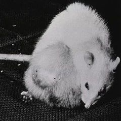
新研究证实细胞移植治疗可恢复盲鼠视力
在一项新的研究中,来自英国牛津大学和新加坡国立大学医院的研究究人员发现,将发育中的细胞移植到盲鼠的眼睛中之后,它们能够再次形成视网膜的整个光敏感层,结果这些小鼠再次能够看见东西。相关研究结果于2013年1月3日在线发表在PNAS期刊上,论文标题为“Reversal of end-stage retinal degeneration and restoration of visual function by photoreceptor transplantation”。论文通信作者为牛津大学纳菲尔德临床神经科学系教授Robert MacLaren。论文第一作者为新加坡国立大学医院眼科外科医生Mandeep Singh博士。
研究人员说,这种方法有望治疗患有色素性视网膜炎(retinitis pigmentosa)的病人。在这种疾病中,视网膜中的感光细胞逐渐死亡从而导致渐进性失明。
研究人员利用由于视网膜中光敏感性的感光细胞(photoreceptor cell)完全缺失而失明的小鼠开展研究。对于治疗因患上色素性视网膜炎而失明的病人而言,它们是最为相关联的模式小鼠。
在两周之后,研究人员发现移植到小鼠眼睛中的细胞在它们的视网膜上再次形成完整的光敏感层。所使用的细胞是处于发育成视网膜细胞的初始过程中的小鼠前体细胞。
瞳孔收缩测试表明,在接受细胞移植的12只小鼠中,有10只小鼠在对光线作出反应时产生的瞳孔收缩得到改善。这就表明这些小鼠的视网膜再次具有感光功能,并且这能够沿着视神经传送到大脑中。
Singh博士说:“我们发现如果一起移植足够多的细胞的话,那么它们不仅是对光线敏感的,而且也能够再生真正意义上的视力所需的连接。”
MacLaren教授解释道:“人们已开展过利用干细胞在病人体内替换视网膜的色素内层(pigmented lining)的临床试验,然而这项新的研究证实感光层可能也以类似的方式被替换掉。这些感光细胞拥有高度复杂的结果。我们观察到在移植到完全失明的视网膜中之后,它们能够作为一个细胞层恢复功能,并恢复连接。”
MacLaren教授解释道,通过展望利用潜在的细胞疗法治疗失明的人们,他们将想要利用诱导性多能干细胞(induced pluripotent stem cells, iPS细胞)开展实验。这些干细胞可以通过重编程病人自己的细胞如皮肤细胞或血细胞而获得,然后经诱导后分化为视网膜细胞的前体细胞。
MacLaren教授说,这点已被其他人成功实现过,因此就目前而言,所要做的就是未来在病人体内开展细胞移植实验。下一步就是在病人体内找到一种可靠的细胞来源,这样就能够提供干细胞用于这些移植实验中。他说,尽管这些是更为长期的开发目标,但是这项研究证实他们可能开发出一种基于细胞的方法。
他说:“我们已证实移植的细胞能够存活下来,它们变成光敏感性的,它们再次形成连接并且形成视网膜的其他部门形成通路从而恢复视力。利用细胞移植重建视网膜的整个光敏感层是干细胞治疗失明的最终目标,这也是我们正在努力实现的。”

 Reversal of end-stage retinal degeneration and restoration of visual function by photoreceptor transplantation
Mandeep S. Singha et al.
One strategy to restore vision in retinitis pigmentosa and age-related macular degeneration is cell replacement. Typically, patients lose vision when the outer retinal photoreceptor layer is lost, and so the therapeutic goal would be to restore vision at this stage of disease. It is not currently known if a degenerate retina lacking the outer nuclear layer of photoreceptor cells would allow the survival, maturation, and reconnection of replacement photoreceptors, as prior studies used hosts with a preexisting outer nuclear layer at the time of treatment. Here, using a murine model of severe human retinitis pigmentosa at a stage when no host rod cells remain, we show that transplanted rod precursors can reform an anatomically distinct and appropriately polarized outer nuclear layer. A trilaminar organization was returned to rd1 hosts that had only two retinal layers before treatment. The newly introduced precursors were able to resume their developmental program in the degenerate host niche to become mature rods with light-sensitive outer segments, reconnecting with host neurons downstream. Visual function, assayed in the same animals before and after transplantation, was restored in animals with zero rod function at baseline. These observations suggest that a cell therapy approach may reconstitute a light-sensitive cell layer de novo and hence repair a structurally damaged visual circuit. Rather than placing discrete photoreceptors among preexisting host outer retinal cells, total photoreceptor layer reconstruction may provide a clinically relevant model to investigate cell-based strategies for retinal repair.


