既往研究发现,狗经过训练能够嗅出“癌症”。
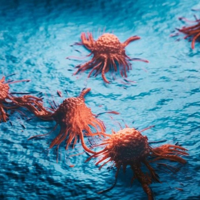
几年前就有报道说宠物狗能嗅出“癌症患者”,最近,一个由马萨诸塞大学阿默斯特分校化学家领导的研究小组开发出一种快速、灵敏的“化学鼻”,能从微观水平识别出活组织内各种细胞类型,几分钟内就能区分出癌转移组织和正常组织。研究人员指出,这为快速诊断癌症提供了一种比较通用的方法,并能减小活体检查的入侵性。相关论文发表在最近出版的《美国化学协会·纳米》杂志上。
迄今为止,精确识别癌细胞的标准方法是用一种能与癌细胞壁结合的生物受体,但这种方法的缺点是必须事先知道相应受体是什么。新研究中,由该校文森特·罗泰洛领导的研究小组用一种黄金纳米粒子传感器阵列加上绿色荧光蛋白(GFP)造出一种新传感器阵列,只需几分钟就能与癌细胞内特殊蛋白质起反应而被激活,从而给每种癌症标上一个独特的识别标志。
在此前研究中,他们已经开发出一种“化学鼻”——由纳米粒子和聚合物组成的阵列,能区分正常细胞和癌细胞。“我们将这一工具用在组织和器官诊断中,能通过‘闻味’的方法实际探测、识别活动物组织中的转移性肿瘤细胞,‘嗅’出不同的癌症类型。”罗泰洛说。
他们用健康组织和小鼠肿瘤样本,不断调节、修整纳米粒子—GFP传感器阵列,一旦发现了转移性组织,GFP就会发出荧光。研究人员解释表示,调整好的传感器阵列能识别各种健康组织,即使组织只有微小变化,它也能“嗅”出来,极为敏感而且功能强大。罗泰洛说:“就好比两块奶酪,看起来一样但用鼻子能分出来哪块美味可口,哪块是几天前的。我们的‘化学鼻’能分出一个组织样本是否正常,是哪种癌症,而且准确率极高。它能分辨仅有2000个细胞的样本,能大大减小活体检查的入侵性。”
除了灵敏度高,“化学鼻”还能区分低转移和高转移,癌症来自哪个部位,如乳腺、肝、肺和前列腺癌。“这一进展让我们向通用型诊断测试更近了一步。总的来说,这种基于阵列的传感策略有望带来一种显型筛选方法,对各种组织情况进行甄别,区分它们是来自基因变异还是组织分化。”研究人员指出,他们下一步将在人体中测试这种传感器阵列。
与能嗅出“癌症患者”的宠物狗相比,“化学鼻”虽然靠的并不是真正的嗅觉,但却不乏亮点:高灵敏度、区分转移组织和癌症类型。癌症之所以可怕,一则在于它早期的隐匿性,一则因为它善于转移。极强的隐匿性使很多患者错过了治疗的最佳时机;而当患者历尽艰辛以为战胜病魔却被告知癌细胞发生转移时,身心都很难再经受住新一轮的折磨。针对这两方面,“化学鼻”在诊断上都有巨大进步。这样的技术一旦推广普及,对于人类健康绝对是一大福音——前提是一定要养成定期体检的良好习惯。

 Array-Based Sensing of Metastatic Cells and Tissues Using Nanoparticle–Fluorescent Protein Conjugates
Subinoy Rana , Arvind K. Singla , Avinash Bajaj , S. Gokhan Elci , Oscar R. Miranda , Rubul Mout , Bo Yan , Frank R. Jirik , and Vincent M. Rotello
Rapid and sensitive methods of discriminating between healthy tissue and metastases are critical for predicting disease course and designing therapeutic strategies. We report here the use of an array of gold nanoparticle–green fluorescent protein elements to rapidly detect metastatic cancer cells (in minutes), as well as to discriminate between organ-specific metastases and their corresponding normal tissues through their overall intracellular proteome signatures. Metastases established in a new preclinical non-small-cell lung cancer metastasis model in athymic mice were used to provide a challenging and realistic testbed for clinical cancer diagnosis. Full differentiation between the analyte cell/tissue was achieved with as little as 200 ng of intracellular protein (1000 cells) for each nanoparticle, indicating high sensitivity of this sensor array. Notably, the sensor created a distinct fingerprint pattern for the normal and metastatic tumor tissues. Moreover, this array-based approach is unbiased, precluding the requirement of a priori knowledge of the disease biomarkers. Taken together, these studies demonstrate the utility of this sensor for creating fingerprints of cells and tissues in different states and present a generalizable platform for rapid screening amenable to microbiopsy samples.


